UDC 581.8
Khani A.B.1, Kozhamzharova L.S.2, Stamkulov E. A2 Kuanbay Zh.I4., AdmanovaG.B.4
1Scientific and Research Institute of Biological Diseases of Zhetysu University named after I. Zhansugurov; Republic of Kazakhstan*
2 International Taraz Innovative Institute named after Sherkhan Murtaza; Republic of Kazakhstan
3,4Aktobe Regional University named after K. Zhubanov; Republic of Kazakhstan
(E-mail: arailim.khani@mail.ru )
Comparative analysis of the anatomical structure of Picea Schrenkiana of the Dzungarian Alatau
Abstract
The morpho-anatomical structure of the leaf used in the diagnosis and taxonomy of species is also a key indicator of its functional characteristics. It is no less important when studying the mechanisms of their adaptation during introduction into new growing conditions and allows us to find ways to optimize the conditions of their vital activity and cultivation. Introduction experience is a necessary condition for studying the adaptive potential of species and for developing methods for managing the growth and development of these plants. Coniferous plants have an invaluable economic value, mainly as raw materials for the production of wood and paper, as well as many conifers are of great importance in gardening and as ornamental garden plants. Despite the above advantages, there is currently a sharp decline in conifers in natural ecosystems. In this regard, the preservation of plant gene pool and rational use of plant resources is an urgent scientific problem. In addition, rooting of coniferous cuttings by the traditional method of reproduction is often very long, from a few months to a year and a half, which is ineffective in restoring the biodiversity of coniferous plants.The genus Picea is represented by many species growing in different geographical regions and not the same ecological conditions. From earlier studies, it can be seen that such a spread significantly affects both the shape of the cells of the needles of the species of the genus Picea, and the size, thickness of the cell walls and mutual arrangement [2, 5, 8]. In this paper, the author continues the earlier study of the comparative anatomy of Picea leaf tissues on the example of P. schrenkiana and P. Pungens. P. schrenkiana or Schrenka spruce is a native of Tien Shan, a tall tree, on average, 40-45 meters tall and up to 2 meters in diameter. The branches are low drooping. The crown is narrowly conical, the branches are low drooping. Needles are thick, hard and sharp, large, thick - about 20-40 mm long, hard and sharp.The color of the needles is bluish-blue, the life span is from 12 to 28 years. P. Pungens, also known as the prickly spruce, is thinner than the Schrenka spruce - the thickness of the trunk reaches only 1.2 meters, the height is about the same - about 45 meters. The crown is cone-shaped, also low-hanging. The needles are coarse, hard, 20-30 mm long, bluish-blue, rarely golden. It keeps on shoots much less than that of the Shrenka spruce - only 4-7 years [1,3,5].
Key words: P. Shrenkiana, cell tissues, slices, coniferous genera, analysis.
Material and methodology
The object of the study was the needles of adult trees Picea schrenkiana and Picea pungens. Conifers were selected from the lower parts of the tree tops from trees grown in the Almaty-Piсea Schrenkiana nature reserve and trees grown in the park of the arboretum of neighboring countries-pikea pungens. The structure of the needles' tissues was studied on preparations fixed in a mixture of Gammalunda, on transverse, paradermal and radial sections prepared according to generally accepted methods. The sizes of the needles' cells were calculated in microns. The data were processed statistically by generally accepted methods. To study the epidermal complex of the leaf, the epidermis was removed using a safety razor blade, and the method of epidermis prints according to J.N. and N.A. Aneli. Anatomical studies were carried out according
to a generally accepted technique with its own modifications on fixed and fresh material using light microscopes P-15, C-11, Nikon ECLIPSE 50i -5 in transmitted light. The general picture of the structure, histological composition, tissues on different sections, and their parameters were established. Measurements were made using a screw eyepiece micrometer MOV 1-15.The sizes of the elements were given in the range "from" and "to" (for example, the diameter of the vessels varies between 100-150 microns) [10,12].
The results of the study
As shown from Table 1, P. the length of the schrenkiana needles corresponds to the data in the examined etiquettes, and P. pungens have slightly shorter needles. The number of layers of P. schrenkiana cells is less than that of P. pungens, both in genuine and in short radius. The indicators showed similar results during the examination.
Table 1.
Parameters of needles Picea schrenkiana and Picea pungens, microns.
|
Indicator
|
Number of cell layers |
Number of cell layers |
||
|
Picea schrenkiana |
Picea pungens |
Piceaschrenkiana |
Picea pungens |
|
|
Long radius |
5,4 ± 0,19 3,0 -7,0 |
8,6±0,22 6,0-11,0 |
22,3 ± 0,95 12,0-31,0 |
17,6 ± 0,39 14,0-22,0 |
|
Short |
3,3 ± 0,19 2,0 -6,0 |
6,2±0,19 4,0 -8,0 |
||
Note. Here and further: in the numerator - average values, in the denominator - the spread of data
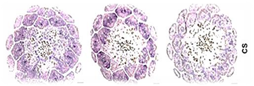
is the long diameter of the needles of P. schrenkiana, approximately corresponds to the short one, that is, the needles have a cross-section close to the square, in P. pungens the leaf is flatter - the short diameter is 1.5 times smaller than the long one.
Ficture 1
Microscopic image of a cross-section of needles (the picture was taken on a Nikon ECLIPSE 50i microscope, size from x10 to x50)

Table 2.
Parameters of the cross-section of needles Picea schrenkiana and Picea pungens, microns.
|
Indicator |
Picea schrenkiana |
Picea pungens |
|
Long diameter |
700,7 ± 26,6 |
1155,0 ± 32,80 |
|
|
387,5 - 1116,0 |
924,0 - 1694,0 |
|
Short diameter |
749,9 ± 33,01 |
882,4 ± 27,41 |
|
|
434,0 - 1240,0 |
646,8-1278,2 |
As can be seen from Table 3, on the cross-section, the epidermal cells of P. schrenkiana and P. pungens differ quite a lot in shape: P. schrenkiana cells are more elongated, almost oval, the cell wall is thinner. P. pungens cells tend to be squarer in shape. The longitudinal section shows that in both species the shape of the cells is elongated, and the surface of the cell membrane is wavy.
Ficture 2
Microscopic image of the parameters of the cells of the epidermis of needles(the picture was taken on a Nikon ECLIPSE 50i microscope, size x50)

Table 3.
Parameters of the epidermis cells of needles Picea schrenkiana and Picea pungens, microns.
|
Cell Sizes |
Picea schrenkiana |
Picea pungens |
|
Length |
54,2 ± 3,13 25,2 - 100,8 |
43,9 ± 1,98 21,6-68,4 |
|
Width |
19,9 ± 0,75 10,8-28,8 |
21,9 ± 0,57 14,4-28,8 |
|
Thickness |
19,9 ± 0,61 14,4-28,8 |
19,4 ± 0,54 14,4-25,2 |
|
Thickness of the outer wall of the epidermis |
7,92 ± 0,39 3,6 - 10,8 |
13,3 ± 0,61 7,2 - 18,0 |
Note. The length of the cell wall was measured on the paradermal section, the width, thickness and thickness of the outer wall on the transverse one.
There is no noticeable difference in the parameters of hypodermic cells of the studied species. In both species, the cells are elongated, the length slightly exceeds the width. The spread of values is also almost the same, as can be seen from Table 4.
Ficture 3
Dimensions of hypodermic cells on cross sections of needles(the picture was taken on a Nikon ECLIPSE 50i microscope, size from x10 to x50)

Table 4.
Sizes of hypodermic cells on cross sections of needles Picea schrenkiana
and Picea pungens, microns
|
Cell Sizes |
Picea schrenkiana |
Picea pungens |
|
Length |
20,0 ± 0,72 |
22,6 ± 0,72 |
|
|
14,4 - 32,4 |
10,8 - 28,8 |
|
Width |
14,4 ± 0,57 |
18,0 ± 0,46 |
|
|
10,8 - 21,6 |
10,8 - 21,6 |
Picea pungens endoderm cells are larger than P. schrenkiana cells, although in both species the cell shape is oval and they are strongly elongated in length (the cell length exceeds the width by almost 1.5 times) (Table 5.).
Table 5.
Endoderm cell sizes on cross sections of needles Picea schrenkianaand Picea pungens, microns
|
Cell Sizes |
Picea schrenkiana |
Picea pungens |
|
Length |
39,6 ± 0,97 |
51,4±1,11 |
|
|
28,8 - 50,4 |
39,6 - 64,8 |
|
Width |
17,3 ± 0,82 |
23,0 ± 0,79 |
|
|
7,2 - 28,8 |
18,0 - 32,4 |
From Table 6 P. pungens P along the length of mesophyll cells. similar to schrenkiana cells, but we can conclude that they are expanding. The cell length of both species exceeds the width, but P. in schrenkiana, there is a difference between length and width.
Ficture 4
View of mesophyll cells on paradermal sections of needles (the picture was taken on a Nikon ECLIPSE 50i microscope, size from x10 to x50)

Table 6.
Dimensions of mesophyll cells in the parаdermal parts of needles
Picea schrenkiana and Picea pungens, microns
|
Cell Sizes |
Picea schrenkiana |
Picea pungens |
|
Length |
32,8 ± 1,11 |
34,9 ± 1,15 |
|
|
18,8 - 46,8 |
21,6 - 46,8 |
|
Width |
22,6 ± 0,68 |
30,6 ± 1,04 |
|
|
18,0 - 28,8 |
18,0 - 46,8 |
From the analysis of the data given in Table 7, it can be concluded that both in length and width and thickness of the mesophyll cells of P. pungens needles exceed the size of P. schrenkiana cells, both in the first layer from the epidermis and in the first layer from the endoderm. In shape, the first layer from the epidermis is represented by elongated thin cells that retain an approximately equal ratio of length, width and thickness in both species. [1,2,11]. The first layer of cells from the endoderm is represented in P. schrenkiana, and cells close to a square shape, although the size spread in length is much larger than in width (the length spread is 50.4 microns, while the width is only 25.2). P. pungens cells are longer, wider and thicker. The basis of their shape is not a square, but rather a rectangle.
Table 7.
Comparative characteristics of mesophilic needle cell sizes Picea schrenkiana and Picea pungens, microns
|
Cell Sizes
|
The first layer from the epidermis |
The first layer from the epidermis |
||
|
Piceaschrenkiana |
Piceaungens |
Piceaschrenkiana |
Piceapungens |
|
|
Length |
41,7 ± 1,76 18,0-72,0 |
50,7 ± 1,72 32,4-68,4 |
41,7 ± 1,83 21,6-72,0 |
63,0 ± 2,30 39,6-90,0 |
|
Width |
30,6 ± 1,29 18,0-46,0 |
33,8 ± 1,69 18,0-61,2 |
41,4 ± 1,44 28,8-54,0 |
42,4 ± 1,29 28,8-54,0 |
|
Thickness |
27,0 ± 1,40 18,0-54,0 |
31,6 ± 1,08 18,0-46,8 |
28,0 ± 1,22 18,0-39,6 |
32,4 ± 0,93 18,0-43,2 |
.
Note. Length and width were measured on cross sections, thickness - on radial sections.
Conclusions
In general, the structure of the needles of the two species of the genus Picea differs more quantitatively than qualitatively: the length of the needles of P. pungens is less than that of P. schrenkiana, but the number of cell layers on the cross-section of the prickly spruce exceeds that of P. schrenkiana, both in long and short radius. The cross-section itself is larger in P. pungens, in other words, the needles of this species are thicker and more rigid. Hypodermal is the only one of the studied tissues, in the parameters of the cells of which there are almost no differences in size or shape in the two Picea species. Endoderm cells are also larger in P. pungens. As for the mesophyll, both on the cross-section, and on the paradermal, and on the radial, the cell sizes of P. schrenkiana are inferior to P. pungens, although the difference is not global.
References
1. Bulygin N. E. Dendrology. (1991) 2nd ed., revised. and additional Leningrad: Agroprom-publ. Leningrad branch, 352 p. - (Textbooks and textbooks for higher educational institutions). - 7300 copies. - TSBN 5-10-001679-5.
2. Gamalei Yu.V. Mesophyll (1985) // Atlas of ultrastructure of plant tissues. Petrozavodsk, pp. 97 - 127.
3. Gorchakovsky P.L., Shiyatov S.G. (1985) Phytoindication of environmental conditions and natural processes in high mountains // Moscow, P. 3
4. Garcia D, Collier SA, Byrne ME, Martienssen RA. Specification of leaf polarity in Arabidopsis via the trans-acting siRNA pathway. Curr Biol. 2006;16:933–8.
5. Iwasaki M, Takahashi H, Iwakawa H, Nakagawa A, Ishikawa T, Tanaka H, Matsumura Y, Pekker I, Eshed Y, Vial-Pradel S, et al. Dual regulation of ETTIN (ARF3) gene expression by AS1-AS2, which maintains the DNA methylation level, is involved in stabilization of leaf adaxial-abaxial partitioning in Arabidopsis. Development. 2013;140:1958–69.
6. Franck DH. The morphological interpretation of epiascidiate leaves—An historical perspective. Bot Rev. 1976;42:345–88.
7. Анели Дж.Н., Анели Н.А. Способ получения микроструктурных отпечатков эпидермы различных органов растений // Сообщения АН ГрузССР. Тбилиси, 1986. С. 589–592
8. Yamaguchi T, Tsukaya H. Evolutionary and developmental studies of unifacial leaves in monocots: Juncus as a model system. J Plant Res. 2010; 123:35.
9. Blokhina, N.I. (2000) Analysis of the age variability of anatomical features of Olginskaya larch wood and correlation with the conditions of tree growth / N.I. Blokhina et al. // Structure, properties and quality of wood. mater. III international. simp. - Petrozavodsk, pp. 37-40.
10. Ovesnov S.A., Perevedentseva L.G. (2007) Morphology and anatomy of vegetative organs of higher plants. Guidelines for laboratory work. [in Russian].
11. Stamatakis A. RAxML version 8: a tool for phylogenetic analysis and post-analysis of large phylogenies. Bioinformatics. 2014;30:1312–3.
12. Brewer PB, Heisler MG, Hejátko J, Friml J, Benková E. In situ hybridization for mRNA detection in Arabidopsis tissue sections. Nat Protoc. 2006;1:1462–7.
Хани А.Б.*, Кожамжарова Л.С., Стамкулов Е.А., Адманова Г.Б., Қуанбай Ж.И.
Сравнительный анализ анатомического строения PiceaSchrenkiana Джунгарского Алатау
Хвойные имеют неоценимое хозяйственное значение, главным образом в качестве сырья для производства древесины и бумаги, а также многие хвойные имеют большое значение в озеленении и в качестве декоративных садовых растений. Несмотря на вышеперечисленные преимущества, в настоящее время наблюдается резкое сокращение видов хвойных растений в природных экосистемах. В этой связи актуальной научной проблемой является сохранение генофонда растений и рациональное использование растительных ресурсов. Кроме того, укоренение черенков хвойных при традиционном способе размножения-черенкование часто очень длительное, от нескольких месяцев до полутора лет, что неэффективно для восстановления биоразнообразия хвойных растений. Род Picea представлен многими видами, произрастающими в разных географических регионах и в неодинаковых экологических условиях. Из более ранних исследований видно, что такое распространение существенно влияет как на форму клеток хвои видов рода Picea, так и на размер, толщину клеточных стенок и взаимное расположение [2, 3, 4]. В этой статье автор продолжает более раннее изучение сравнительной анатомии тканей листьев Picea на примере P. schrenkiana и P. Pungens. P. ель шренкиана или Шренка - уроженка Тянь-Шаня, высокое дерево, в среднем 40-45 метров высотой и до 2 метров в диаметре. Ветви низко поникшие. Крона узко-коническая, ветви низко поникшие. Иглы толстые, твердые и острые, крупные, толстые - около 20-40 мм длиной, твердые и острые. Цвет хвои голубовато-голубой, продолжительность жизни от 12 до 28 лет. P. Pungens, также известная как ель колючая, тоньше ели Шренка - толщина ствола достигает всего 1,2 метра, высота примерно такая же - около 45 метров. Крона конусообразная, также низко свисающая. Хвоя грубая, жесткая, длиной 20-30 мм, голубовато-голубая, редко золотистая. Она держится на побегах гораздо меньше, чем у ели Шренка, - всего 4-7 лет [1,5].
Ключевые слова: P. Shrenkiana, клеточные ткани, срезы, роды хвойных, анализ.
Хани А. Б.*, Қожамжарова Л. С., Стамкулов Е.А., Адманова Г.Б., Куанбай Ж.І.
Жоңғар Алатауының Picea Schrenkiana анатомиялық құрылымын салыстырмалы талдау
Қылқан жапырақты өсімдіктер баға жетпес экономикалық құндылыққа ие, негізінен ағаш пен қағаз өндіруге арналған шикізат ретінде, сондай-ақ көптеген қылқан жапырақты өсімдіктер көгалдандыруда және сәндік бақша өсімдіктері ретінде үлкен маңызға ие. Жоғарыда аталған артықшылықтарға қарамастан, қазіргі уақытта табиғи экожүйелерде қылқан жапырақты өсімдіктердің күрт төмендеуі байқалады. Осыған байланысты өсімдіктердің гендік қорын сақтау және өсімдік ресурстарын ұтымды пайдалану өзекті ғылыми проблема болып табылады. Сонымен қатар, дәстүрлі көбею әдісімен қылқан жапырақты шламдардың тамырлануы көбінесе өте ұзақ, бірнеше айдан бір жарым жылға дейін, бұл қылқан жапырақты өсімдіктердің биоәртүрлілігін қалпына келтіруге тиімсіз.Picea тұқымдасы әртүрлі географиялық аймақтарда және тең емес экологиялық жағдайда өсетін көптеген түрлермен ұсынылған. Бұрынғы зерттеулерден бұл таралу Picea тұқымдас түрлердің инелер жасушаларының пішініне де, жасуша қабырғаларының мөлшеріне, қалыңдығына және өзара орналасуына айтарлықтай әсер ететіндігін көруге болады [2, 3, 4]. Бұл мақалада автор P. schrenkiana және P. Pungens мысалында Picea жапырақтары тіндерінің салыстырмалы анатомиясын ертерек зерттеуді жалғастырады. P. шренкиан немесе Шренка шыршасы-Тянь-Шаньның тумасы, биік ағаш, орташа биіктігі 40-45 метр және диаметрі 2 метрге дейін. Бұтақтар төмен құлайды. Тәжі тар конустық,бұтақтары төмен. Инелер қалың, қатты және өткір, үлкен, қалың - ұзындығы шамамен 20-40 мм, қатты және өткір. Инелердің түсі көкшіл-көк, өмір сүру ұзақтығы 12-ден 28 жасқа дейін. P. Pungens, сондай - ақ тікенді шырша деп те аталады, Шренк шыршасынан жұқа - магистральдың қалыңдығы небары 1,2 метрге жетеді, биіктігі шамамен 45 метрге жетеді. Тәжі конус тәрізді, Сонымен қатар төмен ілулі. Инелер өрескел, қатал, ұзындығы 20-30 мм, көкшіл-көк, сирек алтын. Ол Шренк шыршасынан әлдеқайда аз қашу ұстайды-тек 4-7 жыл [1,5].
Кілтті сөздер: P. Shrenkiana, жасуша тіндері, бөлімдер, қылқан жапырақты тұқымдар, талдау.
Скачано с www.znanio.ru
Материалы на данной страницы взяты из открытых источников либо размещены пользователем в соответствии с договором-офертой сайта. Вы можете сообщить о нарушении.